
Thanks to a mouse viewing scenes from "The Matrix," researchers have developed the most extensive functional map of a brain so far—a chart illustrating how 84,000 neurons connect and communicate with each other.
With just a tiny portion of the mouse’s brain, roughly the size of a poppy seed, scientists were able to pinpoint these specific nerve cells and follow their communication pathways across an unexpected 500 million synaptic connections.
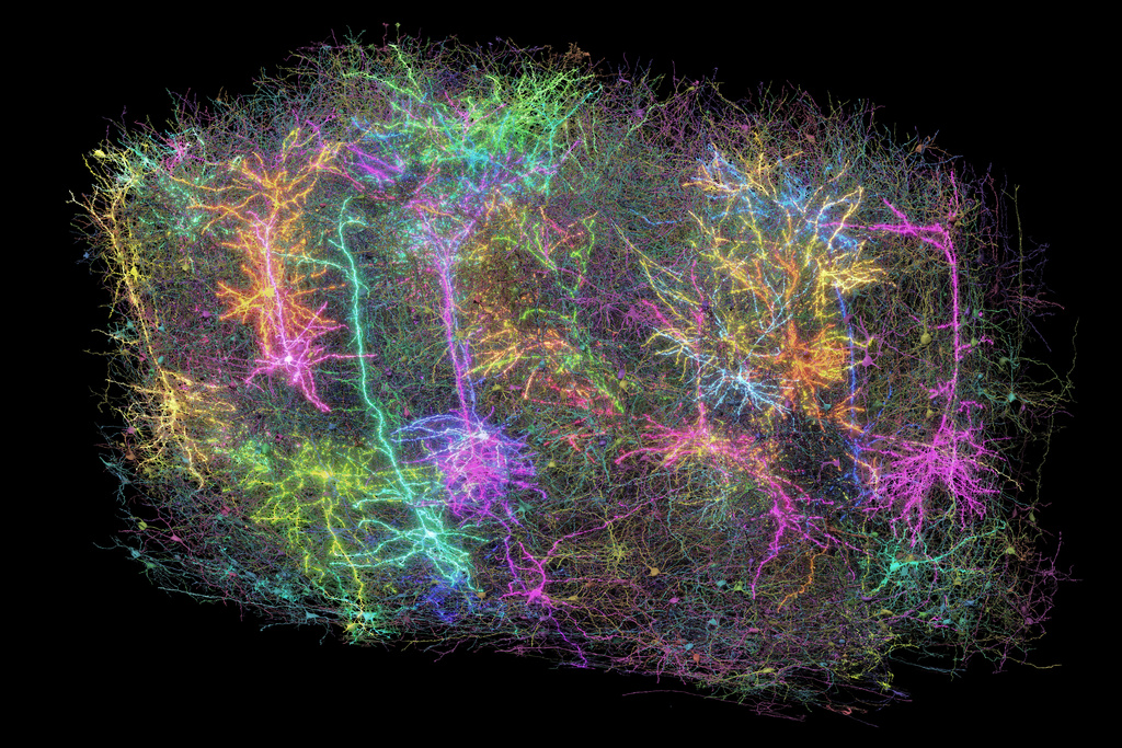
The large dataset, released on Wednesday by the journal Nature, signifies progress in deciphering the enigma of brain function. This information, presented as a three-dimensional recreation with various colors highlighting distinct neural networks, is now accessible to researchers globally for further investigation – and for exploration. simply curious to take a peek.
“Forrest Collman from the Allen Institute for Brain Science in Seattle, a key researcher on this project, mentioned, ‘It instills an overwhelming feeling of wonder similar to viewing images of distant galaxies.’ He added, ‘This makes us realize our own intricacy. Even when examining only a small section of a mouse’s brain, we witness remarkable and complex patterns among thousands of neurons along with their countless interconnections.’”
Our thoughts, emotions, perceptions, speech, and movements result from the activity of neurons—nerve cells—in our brains. These neurons communicate with each other through activation and signaling processes. Experts have understood for some time that these signals travel down structures within individual neurons known as axons and dendrites before being transmitted across gaps called synapses to adjacent neurons. However, much remains unknown regarding the specific neural networks responsible for particular functions and how disturbances in this intricate connectivity might contribute to conditions like Alzheimer's disease, autism, or various other neurological disorders.
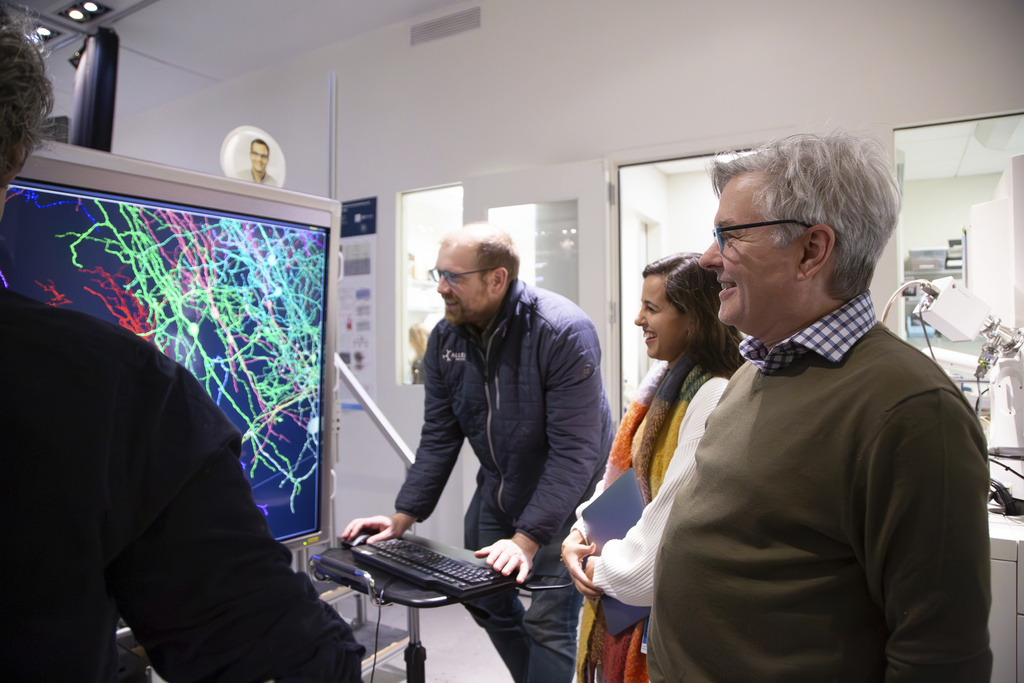
"You could formulate a thousand theories on how brain cells perform their functions, yet without knowing the most basic aspect—the wiring diagram of these cells—you wouldn't be able to test them," explained Clay Reid, an Allen Institute researcher, who was instrumental in advancing electron microscopy for studying neural connections.
For the latest initiative, a worldwide group comprising over 150 scientists charted neuronal pathways within a section of the mouse brain associated with sight. These connections were likened by Collman to strands of spaghetti that have been intricately intertwined.
The initial phase involves presenting brief clips from science fiction films, sporting events, animated shows, and documentaries about nature.
A team at Baylor College of Medicine did just that, using a mouse engineered with a gene that makes its neurons glow when they’re active. The researchers used a laser-powered microscope to record how individual cells in the animal’s visual cortex lit up as they processed the images flashing by.
Next, scientists at the Allen Institute analyzed that small piece of brain tissue, using a special tool to shave it into more than 25,000 layers, each far thinner than a human hair. With electron microscopes, they took nearly 100 million high-resolution images of those sections, illuminating those spaghetti-like fibers and painstakingly reassembling the data in 3D.
Lastly, researchers from Princeton University utilized artificial intelligence to map out all the connections and "assign a distinct color to each wire for individual identification," as Collman stated.
They calculated that if the tiny wires were stretched out, they would extend for over 3 miles (5 kilometers). Crucially, correlating this intricate neural structure with the brain activity of the mouse while it viewed videos enabled scientists to map out the functioning of the neurological circuits.
The Princeton researchers also created digital 3D copies of the data that other scientists can use in developing new studies.
Might such mapping assist scientists in discovering therapies for brain disorders down the line? Researchers view it as an essential milestone, similar to the impact of the Human Genome Project, which offered initial genetic maps leading to gene-specific treatments. Their subsequent aim is to map an entire mouse brain.
"The innovations created through this initiative will provide our initial opportunity to genuinely detect an unusual pattern of connectivity that leads to a disorder," stated Sebastian Seung, one of the project’s key researchers and a neuroscientist and computer scientist from Princeton University.
The work "represents a significant advancement and provides an essential community resource for forthcoming discoveries," noted Harvard neuroscientists Mariela Petkova and Gregor Schuhknecht, who were not part of the project.
The extensive and collectively accessible data "will aid in deciphering the intricate neurological networks that underlie thought processes and actions," they further noted.
The MICrONS consortium, which stands for Machine Intelligence from Cortical Networks, received funding through the BRAIN Initiative at the National Institutes of Health as well as via IARPA, the Intelligence Advanced Research Projects Activity.
—-
The Associated Press' Health and Science Division is supported by the Howard Hughes Medical Institute’s Science and Educational Media Group along with the Robert Wood Johnson Foundation. However, the AP maintains full responsibility for the content produced.