
Thanks to a mouse viewing scenes from "The Matrix," researchers have developed the most extensive functional map of a brain so far, charting the connections among 84,000 neurons as they transmit signals.
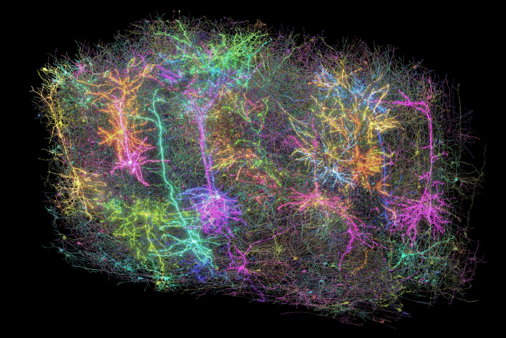
Using a piece of that mouse’s brain about the size of a poppy seed, the researchers identified those neurons and traced how they communicated via branch-like fibers through a surprising 500 million junctions called synapses.
The massive dataset, published Wednesday by the journal Nature, marks a step toward unraveling the mystery of how our brains work. The data, assembled in a 3D reconstruction colored to delineate different brain circuitry, is open to scientists worldwide for additional research – and for the simply curious to take a peek.
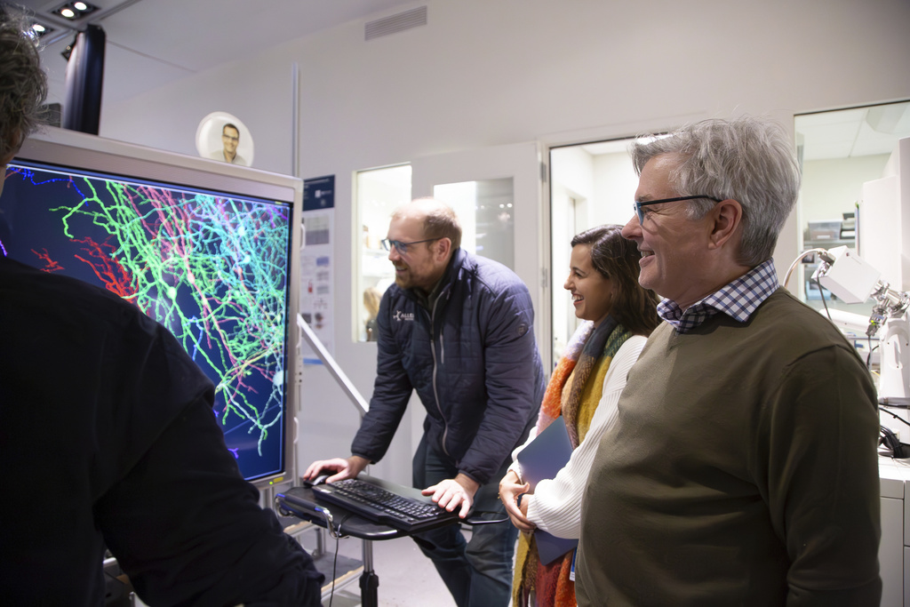
"Forrest Collman from the Allen Institute for Brain Science in Seattle, a key researcher on this project, mentioned, 'It instills a feeling of wonder similar to viewing images of distant galaxies. It makes us realize our intricate nature. We're examining only a small section... of a mouse's brain, yet the elegance and intricacy evident in these individual neurons along with their billions of interconnections are astounding,' " he explained.
Our thoughts, emotions, perceptions, speech, and movements result from the activity of neurons—nerve cells—in our brains. These neurons communicate with each other through activation and signaling processes. Experts have extensively studied how these signals travel down structures like axons and dendrites within individual neurons before crossing over to adjacent neurons via synapses. However, much remains unknown regarding the specific neural networks responsible for particular functions and what might happen when their connections malfunction, potentially contributing to conditions such as Alzheimer's disease, autism, or various other ailments.
"You could formulate a thousand theories on how brain cells function, yet without understanding what may be the most basic aspect—how these cells are interconnected—you won't be able to validate those theories," explained Clay Reid, an Allen Institute researcher, who was instrumental in advancing electron microscopy for studying neural connections.
With the new project, a global team of more than 150 researchers mapped neural connections that Collman compares to tangled pieces of spaghetti winding through part of the mouse brain responsible for vision.
Step one involves displaying short clips from various genres including science fiction films, athletics, animated content, and natural scenes featuring mice.
Researchers at Baylor College of Medicine achieved this by employing a genetically modified mouse whose neurons emit light when activated. They utilized a laser-scanning microscope to capture footage of single cells within the creature’s visual cortex lighting up as they interpreted rapidly displayed visuals.
Following this, researchers from the Allen Institute examined a tiny section of brain tissue by slicing it into over 25,000 slices with an advanced instrument, making each slice much finer than a strand of hair. Using electron microscopes, they captured almost 100 million high-definition photographs of these segments, thereby revealing the tangled network of fibers and meticulously reconstructing the information in three dimensions.
Ultimately, researchers from Princeton University employed artificial intelligence to map out all the connections and "assign a distinct color to each wire for individual identification," as Collman described.
They calculated that if the tiny wires were stretched out, they would extend for over 3 miles (5 kilometers). Crucially, correlating all this anatomical information with the brain activity of the mouse while it watched videos enabled scientists to map out how the neural circuits functioned.
The research team from Princeton also generated digital 3D replicas of the information for other scientists to utilize in their new investigations.
Might such a map assist scientists in developing therapies for brain disorders down the line? Researchers view it as an essential milestone, similar to the impact of the Human Genome Project, which offered initial genetic sequencing and paved the way for gene-specific treatments. Their subsequent aim is to chart an entire mouse brain.
"The innovations created through this initiative will provide our initial opportunity to genuinely detect certain unusual patterns of connectivity that lead to disorders," stated Sebastian Seung, a prominent researcher from Princeton specializing in neuroscience and computer science, as part of his official commentary.
The work "represents a significant advancement and provides an indispensable community resource for forthcoming discoveries," noted Harvard neuroscientists Mariela Petkova and Gregor Schuhknecht, who were not part of the project.
The massive and openly accessible data "will aid in deciphering the intricate neurological networks that underlie thought and action," they further noted.
The MICrONS consortium, which stands for Machine Intelligence from Cortical Networks, received funding through the BRAIN Initiative at the National Institutes of Health as well as from IARPA, the Intelligence Advanced Research Projects Activity.
—-
The Associated Press' Health and Science Division is supported by the Howard Hughes Medical Institute’s Science and Educational Media Group along with the Robert Wood Johnson Foundation. However, the AP maintains full responsibility for the content produced.