
Following nine years of meticulous effort, an international group of scientists released a detailed map of the visual areas in a mouse’s brain on Wednesday. This unveiling exposes the intricate architectures and operational networks responsible for mammalian sensory processing.
To date, it is the largest and most detailed such rendering of neural circuits in a mammalian brain.
The map aims to expedite research into how healthy brains operate: perceiving, retaining and managing memories, and maneuvering through intricate spaces. Equally significant, it will enhance our understanding of neurological disorders from an anatomical and physiological perspective—specifically, in relation to neural connections and signal interactions. This advancement holds particular promise for conditions potentially stemming from irregular circuitry, like autism and schizophrenia.
The innovations created through this initiative will provide our initial opportunity to genuinely pinpoint an unusual connectional pattern that could lead to a disorder," stated H. Sebastian Seung, the Evnin Professor in Neuroscience and a professor of computer science at Princeton University, who jointly led the project.
The work was released as part of a series of papers making up a special issue of the journal Nature .
In addition to Seung's group at Princeton, the consortium was jointly led by teams from the Baylor College of Medicine and the Allen Institute for Brain Science. The initiative involved over 150 scientists from 22 different organizations.
The NIH officials have pointed out that this research serves as a cornerstone for upcoming investigations into well-being, illness, and abnormal conditions. The agency responsible for gathering information has expressed an aim to deconstruct the mental processes of the brain with the intention of applying these insights to advanced artificial intelligence methods. For instance, mammal brains excel at utilizing limited data to reach intricate conclusions compared to computer systems. This chart introduces a novel approach to examining their capabilities.
Aligning form with function within the brain
As this biological material within our skulls enables us to perceive the world, experience emotions, and make choices," explained Andreas Tolias, a neuroscientist who led this initiative alongside others. He was previously affiliated with Baylor but has since moved to Stanford University.
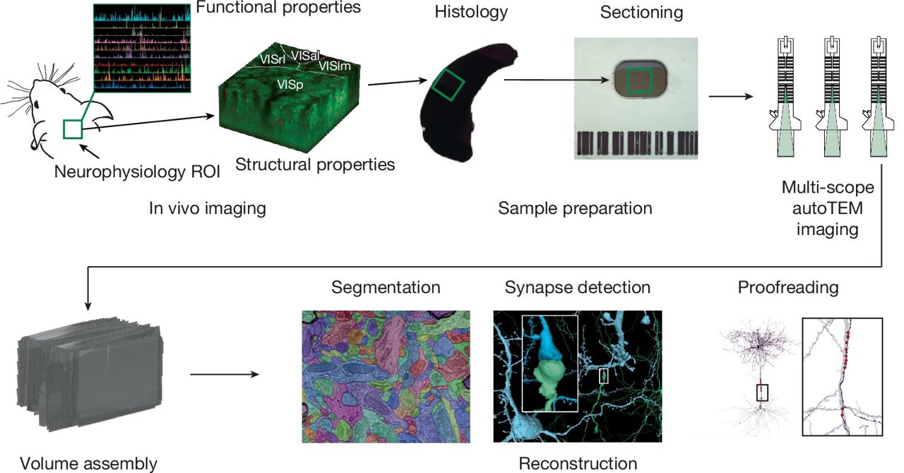
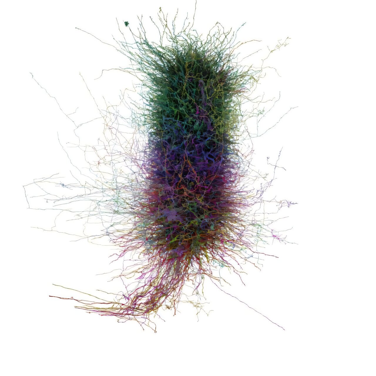
When creating the map, the researchers digitally untangled tens of thousands of individual neuronal cells shaped like trees. They followed each neuron’s unique branching structure and subsequently rebuilt them individually into an extensive neural network—the connectome as it is known among scientists.
The outcome encompasses over half a billion connections within a single cubic millimeter of brain tissue, covering areas such as the primary visual cortex and the retina.
"The uniqueness of this data lies in bringing both the structure and function together within a single experiment," according to Tolias.
While connected to an advanced imaging setup, the mouse was presented with brief video segments. Using this technology, scientists monitored the creature’s neural activity patterns through changes in calcium ion levels, which reflect how data moves within the brain.
They were interested in understanding both the organization of that activity within the brain as well as how this activity correlates with the fundamental cells and their interconnections.
Walking the path
To obtain this information, the scientists required the mouse to remain conscious and visually engaged. Therefore, they made the rodent run on a treadmill while viewing stimulating 10-second video segments, which included excerpts from movies like "The Matrix," "Mad Max: Fury Road," as well as parts of the experimental documentary series known as the Qatsi trilogy, alongside footage of high-intensity activities such as motocross racing, BASE jumping, and luge competitions.
The mouse was subsequently transferred to the Allen Institute, where scientists divided the brain into approximately 28,000 extremely thin sections. They utilized an electron microscope to capture images of each section before assembling these pictures into a comprehensive whole.
Seung’s team subsequently utilized artificial intelligence to map out each contour of every neuron across these myriad slices, highlighting them distinctly with coloration—a procedure known as segmentation. ThisAI-driven segmentation method needs to be confirmed or meticulously reviewed by human experts. An extensive section of the diagram has undergone this verification step; however, the endeavor remains ongoing.
The combined insight into both the structure of the connectome and its functionality renders this technology highly potent. Researchers within the consortium have uncovered several unexpected discoveries regarding neuronal connections, the distinct inclinations of particular neuron classes, as well as the impact these characteristics exert upon complex cognitive processes.
"I believe these issues are merely scratching the surface," Tolias stated.
They have utilized data from this initiative to generate highly detailed digital replicas of the mouse brain, referred to as digital twins, which can be employed to investigate novel inquiries and formulate nuanced hypotheses that can subsequently be confirmed through laboratory experiments.
Thomas Macrina was part of Seung's team at Princeton University as a graduate student, where he obtained his Ph.D. in both neuroscience and computer science during the duration of their work together. Afterward, he founded a company named Zetta AI. This enterprise specializes in handling several intricate aspects of connectome mapping for various research teams—including aligning images, tracking the numerous extensions of neurons, and verifying the accuracy of these findings.
Macrina mentioned that due to advancements made with the mouse project, the techniques have significantly improved in speed and efficiency over recent years. Researchers are currently charting connectomes for various species ranging from mosquitoes and flies to macaques and humans. With these tools becoming increasingly accessible, they hold the potential to expedite findings throughout all areas of neuroscience, allowing scientists to base their investigations into function and behavior firmly within the actualities of neural circuitry.
Macrina stated that each neuroscience experiment should somehow make reference to a connectome.
Achieving 'the impossible'
Writing in Scientific American In 1979, the preeminent biologist of his time, Francis Crick, advised neuroscientists working on technology development to concentrate on objectives within reach. He stated, “Asking for the unachievable isn’t helpful; consider trying to obtain an accurate map of every neuron’s connections in just one cubic millimeter of brain tissue along with their respective activities.”
His words served as a challenge.
Seung drew parallels between the extensive effects of charting the human connectome and how the Human Genome Project revolutionized genomics. Prior to the completion of this project in 2003, humans lacked fundamental knowledge such as gene count per individual and genetic similarities among different individuals.
Twenty years later, genomics has become central to medical practice, allowing tailored therapies for conditions like breast cancer and leukemia, as well as aiding physicians in adjusting the dosage of blood-thinning medications.
Seung stated that the connectome marks the start of the digital revolution in neuroscience. Now, with just a few key presses, one can retrieve information within seconds that previously might have required an entire doctoral dissertation to obtain. This exemplifies the immense potential of this digital shift.
Certainly, significant distinctions exist between the genome and the connectome. Specifically, while the genome can be expressed with a one-line sequence utilizing a four-letter code, the brain consists of intricate neural networks processing data promptly under stringent energy constraints.
While the transformation of brain science could prove to be even more breathtaking than that of genomics, it will also take more effort and creativity to pull off.
Tolias stated, "The connectome does not inherently represent the neural code." He emphasized that although comprehending the intricate details of the brain’s structure is crucial for grasping its functionality and vital for investigating diseases, addressing major queries related to attention and cognitive processes will also necessitate a profound comprehension of the brain's operational principles.
From nematode reproduction to human consciousness
Beginning with modest steps has empowered researchers to develop an array of methods and tools that have helped them progress towards studying larger and more intricate neural structures. In 2019, the full connectomes for both mature males and females of the nematode worm C. elegans were finalized. Then, last year, a group led by Sebastian Seung at Princeton unveiled the entire connectome of a fruit fly.
A number of the researchers involved in that project also contributed to the work with mice. Actually, many of the crucial methodologies that were created and improved for studying one species were applied to investigate the other as well, and conversely.
The data set for the mouse brain is completely accessible to the public, which means scientists worldwide have begun utilizing it to examine hypotheses and devise innovative approaches to understand the brain.
A single cubic millimeter of a mouse’s brain is approximately 20 times larger and significantly more intricate compared to an entire fruit fly brain. However, this fresh mapping effort still falls short of full representation as it covers merely one thousandth of the total mouse connectome.
Transitioning from an incomplete mouse brain connectivity map to a comprehensive human one requires significant time, funding, and creativity—often making such progress appear unattainable. However, what seems improbable today might become reality tomorrow; after all, even the present maps once looked out of reach not too long ago. In fact, as stated by the scientists involved since 2016, their endeavor was akin to Francis Crick’s perspective at that point.
The main takeaway from this project is evidence that constructing connectomes is indeed worthwhile. J. Alexander Bae, who holds a joint Ph.D. in electrical engineering and neuroscience and has been working on this initiative for decades, noted that the concept of charting hundreds of thousands of cells and integrating them into a comprehensive 3D model was quite daring back then.
They encountered significant doubt from their scientific peers. Much of the strenuous effort was carried out manually. Additionally, they had to develop numerous tools required to finish the task.
“It was agonizing. Yet, against all odds, we succeeded. I’m amazed,” Bae stated. “There was potential for failure. However, should we have faltered, the entire field of connectomics might have crumbled.”
Rather, connectomics is poised for rapid expansion.
As scientists encounter greater sizes and complexities, they develop innovative methods, create advanced technologies, and tackle fresh challenges that lead to even bigger and more intricate outcomes, prompting further investigation.
Just the start," Seung remarked. "However, it’s paving the way for a new epoch of lifelike brain simulations. Consequently, the ensuing query will arise—and individuals will inquire—could such an endeavor ever be accomplished with a human brain? Then comes another question: assuming one could replicate a human brain accurately, would this simulation possess consciousness?
When questioned for his opinion on the matter, he chuckled. "I possess no greater authority to comment on this topic than you do. However, whenever someone asserts, ‘I don’t think a simulated brain could be truly aware,’ my response is, ‘How can you be certain that you yourself aren’t just a simulation?’"
More information: The MICrONS Consortium, which covers functional connectomics across various regions of the mouse visual cortex, Nature (2025). DOI: 10.1038/s41586-025-08790-w
Provided by Princeton University
This tale was initially released on Medical Xpress . Subscribe to our newsletter For the most recent science and technology news updates.