
What started as a tiny piece of brain tissue no bigger than a grain of sand has now led to a scientific accomplishment previously considered unattainable. An international group of scientists has presented the most detailed map yet produced of a mammal’s brain structure.
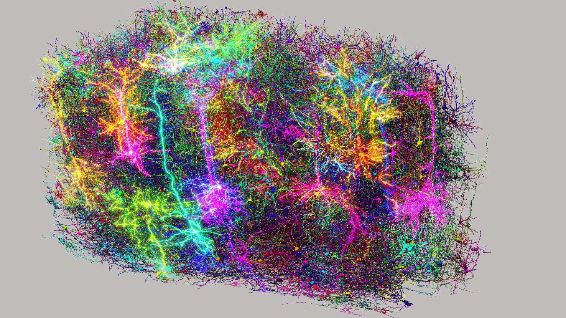
Led by the MICrONS (Machine Intelligence from Cortical Networks) Project, this research offers a detailed, three-dimensional map of a section of a mouse's visual cortex—reshaping our understanding of brain architecture and operation.
The dataset, amounting to an impressive 1.6 petabytes, includes the electrical activity from over 200,000 neurons, 2.5 miles (4 kilometers) of extending dendrites, and over 500 million synapse connections. This online-accessible digital model, available through the MICrONS Explorer, represents a significant milestone for the field of neuroscience.
"The advancements made by MICrONS represent a pivotal point in the field of neuroscience, akin to the impact of the Human Genome Project due to their potential for transformation," stated David A. Markowitz, who coordinated this research effort.
From fragments to synapses: The creation of the brain map
This ambitious project demanded tight cooperation among leading organizations like Princeton University, the Allen Institute, and Baylor College of Medicine. The initiative kicked off when researchers examined neuronal responses in the visual cortex of a mouse as it viewed particular video compilations.
The Allen Institute subsequently examined that section of brain tissue through electron microscopy following the process of cutting it into over 25,000 extremely thin slices, with each slice being finer than a strand of human hair. Experts specializing in artificial intelligence from Princeton applied sophisticated machine learning techniques to map out and rebuild the neural and synaptic connections within the model’s cell structure. They consolidated this information into a comprehensive three-dimensional representation.
Within that minuscule point lies a complete structure akin to a delicate woodland," explained Clay Reid, senior investigator. "This intricate design follows numerous connection principles we've observed across different areas of neuroscientific study. Furthermore, this reconstruction allows us to examine previous hypotheses and strive to uncover novel elements never witnessed before.
One of the most notable findings was an updated understanding of how inhibitory neurons function. Previously thought to solely decrease brain activity, these neurons were found to be quite precise—while some worked together to suppress entire groups of excitatory cells, others targeted almost just one specific kind of cell. This complex system of inhibition and activation unveiled a previously unrecognized equilibrium concerning neuronal coordination, overturning longstanding theories regarding the brain's method of handling information.
Mapping the Future Directions in Neuroscience
This represents the future in numerous aspects," stated Andreas Tolias, one of the principal researchers. "MICrONS will be remembered as a pivotal point where we develop comprehensive brain foundation models that encompass various levels of examination, starting from behavior down to the representation of neural activity and extending all the way to the molecular scale.
In addition to revealing the structure of cognition, this study could pave the way for advancements in identifying and addressing various conditions. brain-related disorders Similar to conditions like Alzheimer’s, autism, and schizophrenia, having a detailed "wiring map" of normal brain tissue allows researchers to start contrasting it with disease models. This process helps them pinpoint where disruptions in signaling happen.
If your radio isn't working and you possess the circuit schematic, you'll be more likely to repair it successfully. said Nuno da Costa, an associate researcher at the Allen Institute, stated, "We're essentially developing a sort of Google map or blueprint for this speck of sand. Going forward, we will be able to utilize this to contrast the neural connections in a healthy mouse with those in another subject." brain wiring within a disease model.
The research has been released in Nature .