
An advancement in imaging tech has the potential to revolutionize our comprehension of how living cells function internally and offer valuable perspectives on numerous ailments.
The research, recently published in the journal Nature Communications unveils a cutting-edge method that merges super-resolution microscopy with AI and deep learning techniques to uncover subcellular details and activities. This research was spearheaded by scientists from Peking University, Ningbo Eastern Institute of Technology, and the University of Technology Sydney.
Imagine flying over a city at nighttime observing all the ongoing activities below," stated UTS Distinguished Professor Dayong Jin. "This advanced technology will unlock new pathways in our endeavor to comprehend the complex universe inside our cells.
A multitude of illnesses and health issues stem from irregularities at the cellular level. Through the visualization of these cellular activities, researchers can gain deeper insights into the fundamental origins of ailments such as cancer, neurodegenerative disorders, and metabolic syndromes, paving the way for enhanced therapeutic approaches.
This novel method addresses several major issues associated with present imaging tools utilized for visualizing components inside living cells.
"As stated by Professor Jin, who leads the UTS Institute for Biomedical Materials & Devices, current technologies like fluorescence microscopy struggle with resolution limits, making it challenging to observe minute cell structures or closely follow intricate cellular activities," he explained.
Conventional techniques may lead to phototoxicity and photobleaching—cell damage from light exposure—and they often find it challenging to concurrently display various structures inside a cell because of limitations in the color palette available.
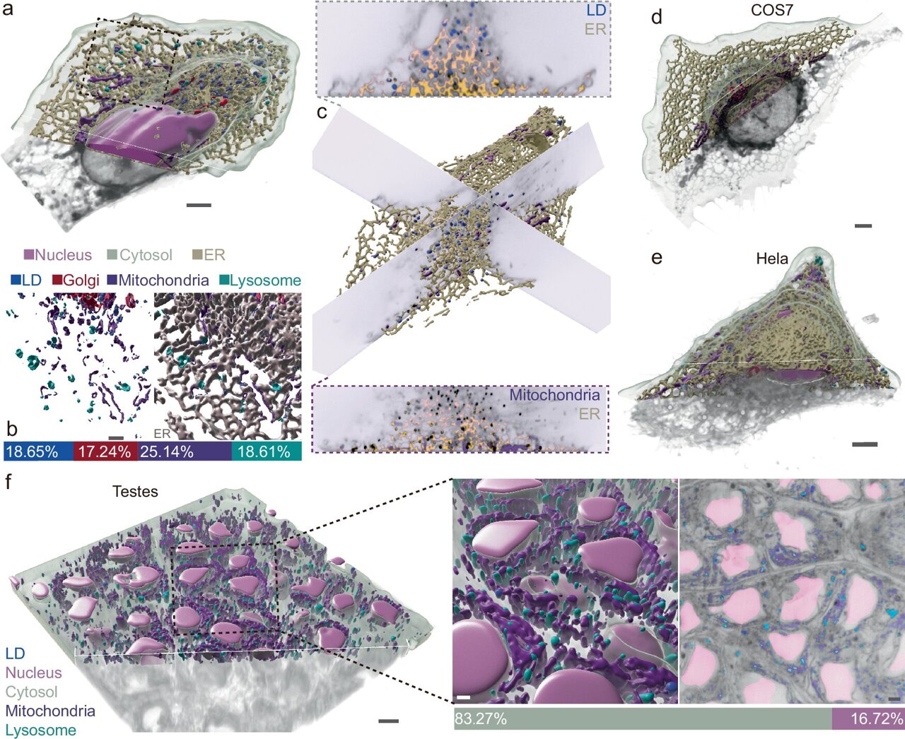
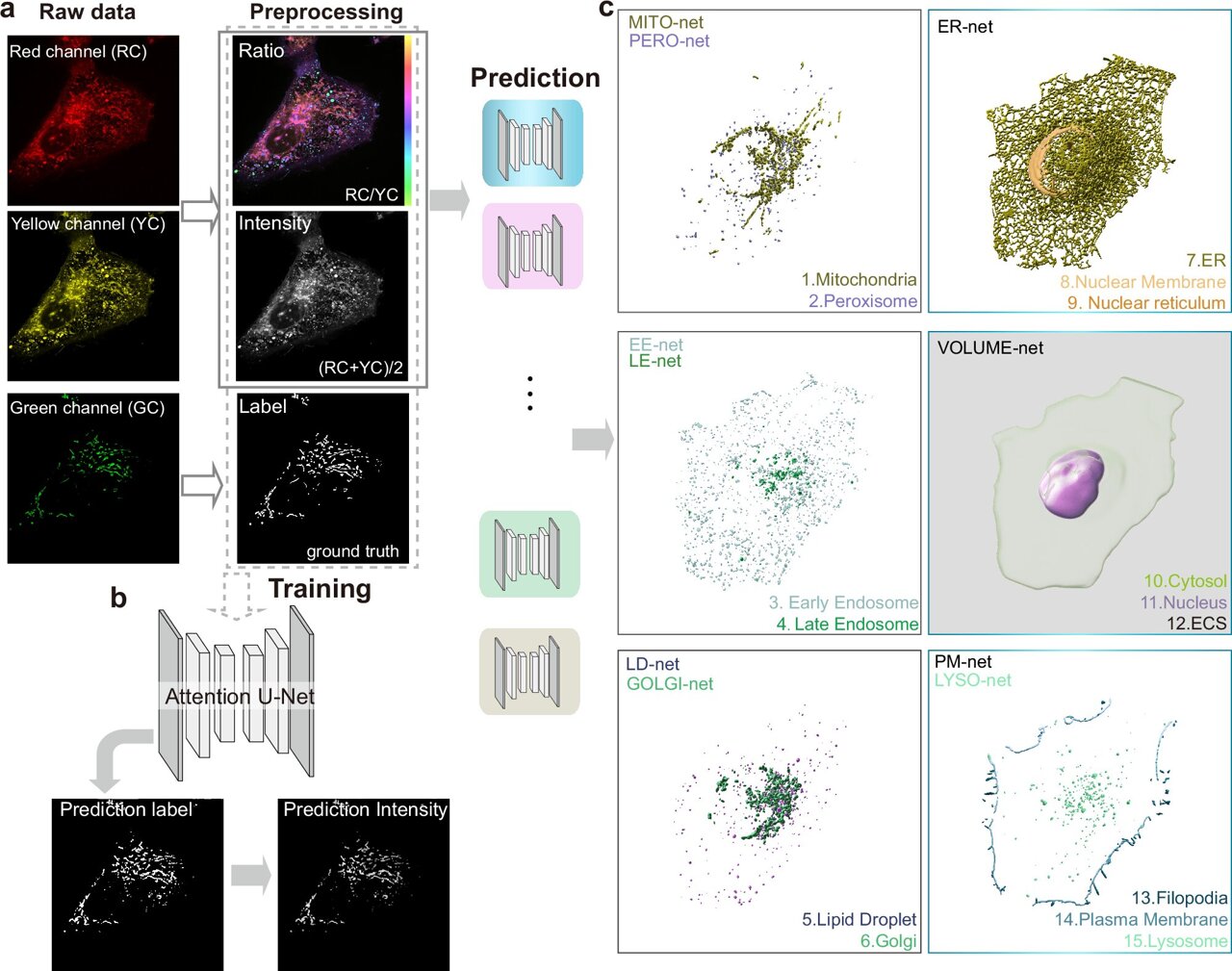
The novel technique can forecast 15 distinct subcellular structures with great precision by employing only one laser and two detection channels.
This advancement doesn’t just overcome the limitations of employing various hues with a singular dye marker; it also greatly accelerates the imaging procedure.
High-quality images precisely depict the distinctions among organelles (cell compartments), functioning akin to an "optical fingerprint." This technique is remarkably versatile and can be utilized with different microscopes, cell varieties, and intricate living tissues.
This flexibility enables researchers to investigate and comprehend the three-dimensional architecture of living cells throughout various phases of cell division and witness swift interplays between intra-cellular sections.
Professor Jin mentioned that the team is collaborating with several medical research institutions at present. This includes virologists who study how viruses interact with cells and the defenses cells employ against them, as well as researchers using advanced imaging techniques to examine cardiomyocytes for deeper understanding of heart conditions. The aim is for this technological approach to yield fresh perspectives and advancements within the field of medical research.
More information: Karl Zhanghao along with colleagues presented an approach for rapid segmentation and multiple image capture of cellular organelles in living cells. Nature Communications (2025). DOI: 10.1038/s41467-025-57877-5
Furnished by the University of Technology, Sydney
This tale was initially released on Massima . Subscribe to our newsletter For the most recent science and technology news updates.