
Thanks to a mouse observing segments from "The Matrix," researchers have developed the most extensive functional brain map to date—a chart illustrating how 84,000 neurons connect as they transmit signals.
Thanks to a mouse observing snippets from "The Matrix," researchers have produced the most extensive functional brain map to date—a chart illustrating the connections between 84,000 neurons as they transmit signals.
With just a tiny section of the mouse’s brain, roughly the size of a poppy seed, scientists were able to pinpoint these specific nerve cells and follow their communication pathways as they extended surprisingly across 500 million connection points known as synapses.
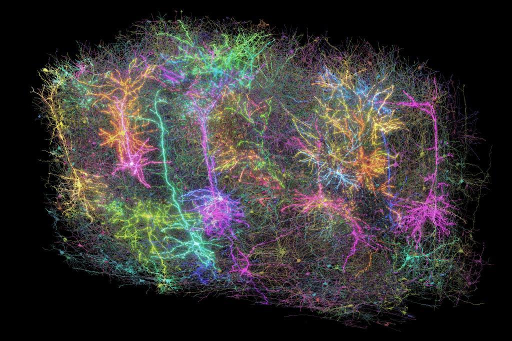
The extensive dataset, released on Wednesday in the journal Nature, represents progress towards understanding the complexities of brain function. This information, presented as a three-dimensional reconstruction with various colors indicating distinct neural pathways, is available globally for researchers and anyone interested to explore further.
Forrest Collman from the Allen Institute for Brain Science in Seattle, a key researcher involved with this initiative, mentioned: 'It certainly evokes feelings of wonder similar to viewing images of distant galaxies,'” he explained. “‘This makes us realize our intricate nature. Even when we examine merely a small segment... of a mouse’s brain, the elegance and intricacy present within individual neurons as well as their countless interconnections become apparent.'
Our thoughts, emotions, perceptions, speech, and movements all result from the activity of neurons—nerve cells—in our brains. These neurons communicate with each other through activation and signaling processes. Experts have extensively studied how these signals travel down structures like axons and dendrites within individual neurons before crossing over to adjacent neurons via synapses. However, much remains unknown regarding the specific neural networks responsible for particular functions and what impact disturbances in this complex circuitry might have on conditions such as Alzheimer’s disease, autism, or various other neurological disorders.
“You could formulate a thousand theories on how brain cells perform their functions, yet without knowing arguably the most crucial aspect—the wiring of these cells—you won’t be able to test those theories,” explained Clay Reid, an Allen Institute scientist instrumental in pioneering electron microscopy for studying neural connections.
In the new initiative, over 150 scientists from around the world collaborated to chart neuronal pathways within a section of the mouse brain associated with sight. These connections were likened by Collman to strands of spaghetti crisscrossing haphazardly inside this complex region.
The initial phase involves displaying short clips from science fiction films, athletics events, animated content, and documentaries about wildlife to the test subject.
Researchers at Baylor College of Medicine achieved this by employing a genetically modified mouse whose neurons emit light when activated. They utilized a laser-scanning microscope to capture the activity of specific cells within the rodent’s visual cortex as various images were displayed rapidly before them.
Following this, researchers from the Allen Institute examined a tiny portion of brain tissue. They utilized a specialized device to slice it into over 25,000 slices, with each one being much finer than a strand of human hair. Using electron microscopes, they captured almost 100 million high-resolution photographs of these segments, clearly showing the threadlike strands and meticulously reconstructing the information into a three-dimensional model.
Lastly, researchers from Princeton University utilized artificial intelligence to map out all the connections and "assign a distinct color to each wire for individual identification," as Collman pointed out.
They calculated that when extended, the tiny wires would total over 3 miles (5 kilometers) in length. Significantly, correlating all this anatomical data with the brain activity of the mouse while it was watching videos enabled scientists to map out how the neural circuits functioned.
The team from Princeton also generated digital 3D replicas of the information for other researchers to utilize in their own investigations.
Might such a mapping assist scientists in discovering therapies for brain disorders down the line? Researchers view it as an essential milestone, similar to how the Human Genome Project’s initial genetic map paved the way for gene-specific treatments. Their subsequent aim is to complete a comprehensive map of a mouse brain.
The innovations created through this initiative will provide our initial opportunity to genuinely detect an unusual pattern of connectivity that leads to a disorder,” stated Sebastian Seung, a prominent researcher from Princeton specializing in both neuroscience and computer science, as part of the project’s announcement.
"The effort represents significant progress and provides an indispensable communal asset for upcoming breakthroughs," noted Harvard neuroscientists Mariela Petkova and Gregor Schuhknecht, who were not part of this initiative.
The extensive and collectively accessible data "will aid in deciphering the intricate neurological networks responsible for thought processes and actions," they noted.
The MICrONS initiative, which stands for Machine Intelligence from Cortical Networks, received funding through the BRAIN Initiative at the National Institutes of Health as well as from IARPA, known as the Intelligence Advanced Research Projects Activity.
Don't overlook all of our updates and information here. www.mundoamerica.com .